Partial Bite Reconstruction
After Occlusal Splint Therapy and Occlusal Equilibration (Bite Adjustment) have been performed, a Partial Bite Reconstruction may be required to correct an unprotective bite in left, right or forward bites.
Partial Bite Reconstruction – Left, Right and Forward Bites
Occlusal Splint Therapy and Occlusal Equilibration will give you a protective hinge bite, that is balanced on both left and right sides on the back teeth. However, your prosthodontist will inform you if one, two or three of your other bites are unprotected.
For example, an unprotected right bite would mean one of your right canines is too short, and not protecting your teeth or your jaw joints when you slide your jaw to the right side. Your right bite may be unprotected because you have worn your lower right canine tooth away from grinding at night, or it may be on the wrong angle.
To correct this, the tooth may need a composite resin overlay or a crown to create a right protective bite and protect your teeth and jaw joints as shown below.

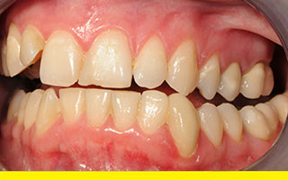
Before – Lower right canine is too short and therefore unprotective
Before – As the lower right canine is too short, it allows the top right lateral incisor to crash into the opposing tooth and the jaw to move directly to the right with ‘cow-like’ movement (Horozonto-Lateral jaw movement) which is not recommended.
Treatment – 0.4mm of the tooth is reduced on the facial side and 1mm on the biting edge. Tooth coloured composite resin filling material is placed as an overlay to build up the canine to its optimal height and function.
After – The lower jaw now moves in a protective manner called Vertico-Lateral jaw movement. This human jaw movement protects the opposite jaw joint, that is, the left TMJ jaw joint and teeth. As the jaw slides to the right the angle and height of the tooth opens the jaw and protects the other teeth from being damaged or prematurely wearing away.
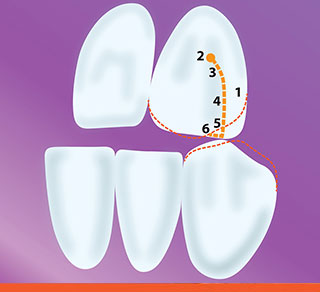
The shape of canine teeth are responsible for the angle the lower jaw moves and the strain placed on the TMJs. The red outline shows the normal anatomy of a canine tooth whereas the white outline of the canines shows a bio-designed tooth shape that creates a left protective bite (Internal view).
For a more detailed explanation on Full Occlusal Protection see Page 5, Figure 5b.
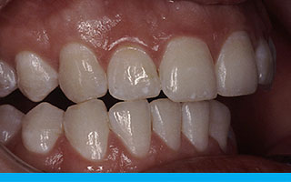
An unprotected forward bite is where your front teeth only touch in one spot or more precisely, in a protected forward bite your two top front teeth should touch together with your lower front teeth simultaneously. An unprotected forward bite will imbalance your jaw and displace your TMJ discs. This concentration of biting force in the wrong area will ultimately chip one of your front teeth.
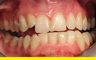
Before – Only one tooth is touching in forward bite and is therefore unprotective to the jaw joints and teeth
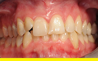
Before – The patient’s lower right canine is angled forward in a damaging manner
Before – The patient’s upper right lateral incisor is pushed forward in a negative manner and the right canines do not touch, allowing the front teeth to be chipped and damaged. This is classified as a right unprotective bite
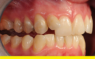
Before – The lower left canine has previously been treated by crowning to create a protective left bite.
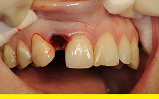
During Treatment – The patient’s crooked tooth has been removed to allow a porcelain bridge to cosmetically replace the crooked tooth. The patient was offered orthodontic therapy but declined due to the cost, time involved and other aesthetic reasons (matching colour and uniform finish). This socket was filled with artificial powdered bone to prevent bone and gum height loss.
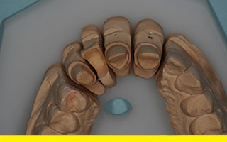
Laboratory model showing the missing tooth with adjacent teeth prepared for a Prettau Zirconia bridge and crown.
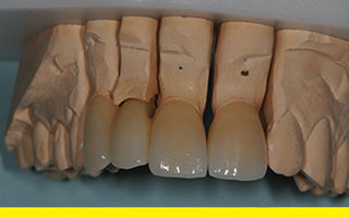
Laboratory model showing the Prettau Zirconia bridge and crown. Porcelain has built-in colours to appear natural.
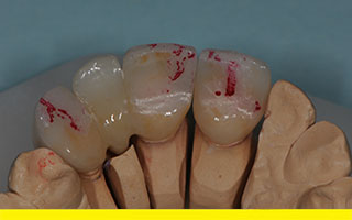
Laboratory model showing the biting view of the Prettau Zirconia bridge and crown with carbon paper marks where the teeth should slide in a protective manner forward and right.
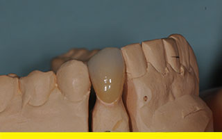
Laboratory model showing the Prettau Zirconia crown for the lower right canine.
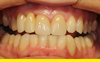
After – Completed natural looking Prettau Zirconia bridge and crowns bio-designed in a protective forward bite.
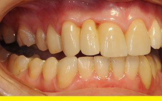
After – Protective right bite.
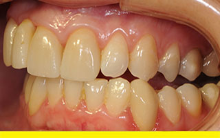
After – Protective left bite.
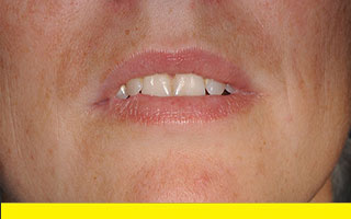
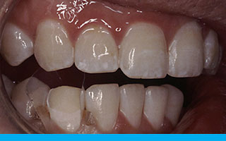
Before – Patient’s lip line showing crooked upper right lateral incisor pinching lip (over exposed photograph).
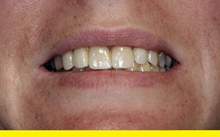
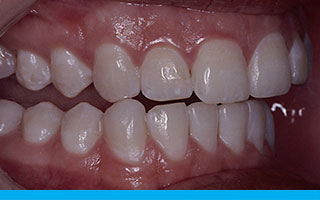
After – Patient’s smile showing new exciting teeth (Slightly under exposed photograph).
Unprotected bites ultimately lead to damaged teeth and prevent full recovery of TMJ symptoms.
Your individual arrangement of teeth will determine how much treatment is required by Your Dental Specialist to achieve all four fully protective bites as demonstrated above with two very different patient cases.